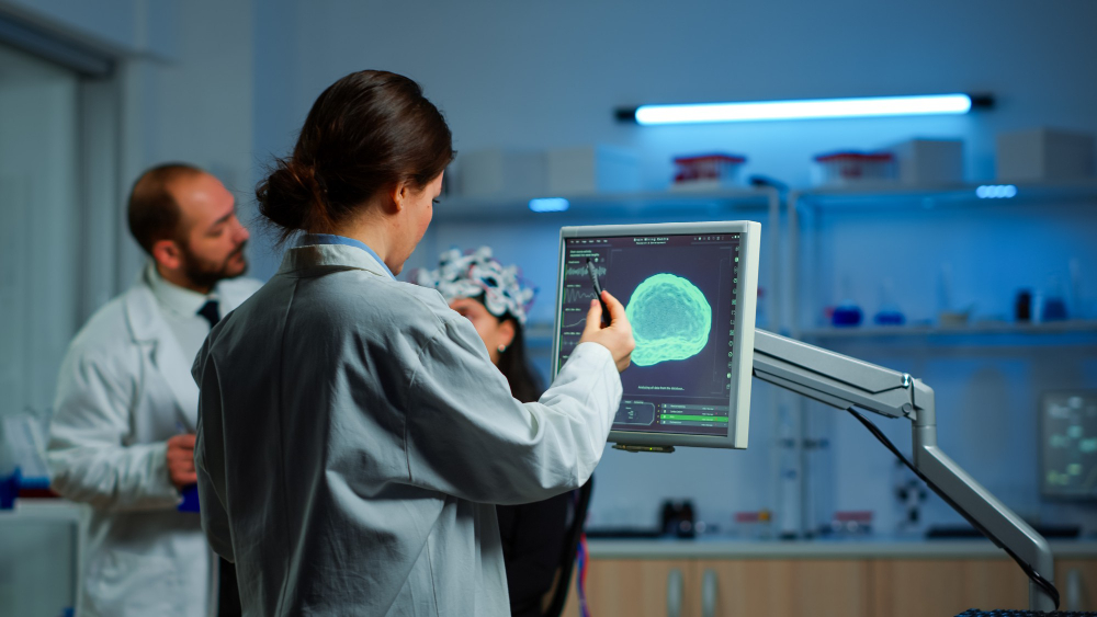
Modern neuroimaging has become one of the most significant advancements in neuroscience, enabling clinicians to diagnose brain diseases with greater speed and precision than ever before. Unlike traditional neurological examinations that rely heavily on symptoms, today's brain imaging technologies provide detailed views of brain anatomy, blood flow, metabolism, and neural activity. This allows physicians to detect diseases such as Alzheimer's disease, Parkinson's disease, epilepsy, stroke, multiple sclerosis, and brain tumors much earlier in their progression. Organizations including the National Institutes of Health (NIH), the National Institute of neurological disorders and Stroke (NINDS), and the Radiological Society of North America (RSNA) continue to highlight the growing role of advanced imaging in improving patient outcomes.
Brain disorders affect millions of people worldwide and often progress silently before noticeable symptoms appear. Detecting these conditions at an early stage can dramatically improve treatment options and patient outcomes. Modern brain imaging technologies allow healthcare professionals to visualize subtle structural and functional changes within the brain, helping them diagnose diseases before irreversible damage occurs.
One of the greatest advantages of modern neuroimaging is its ability to identify neurological disorders at their earliest stages. Diseases such as Alzheimer's disease, Parkinson's disease, multiple sclerosis, and vascular dementia often begin years before patients experience obvious symptoms. Advanced imaging techniques such as high-resolution MRI, diffusion tensor imaging (DTI), and positron emission tomography (PET) can detect microscopic structural and biochemical changes that are invisible through conventional examinations. Early diagnosis enables physicians to begin treatment sooner, recommend appropriate lifestyle modifications, and closely monitor disease progression.
Accurate diagnosis forms the foundation of effective neurological care, and modern brain imaging provides clinicians with highly detailed information that supports every stage of patient management. Instead of relying solely on neurological symptoms, physicians can directly observe abnormalities affecting brain tissue, blood vessels, nerve pathways, and metabolic activity. This detailed visualization helps differentiate conditions that may produce similar symptoms, reducing diagnostic uncertainty and minimizing delays in treatment. Imaging findings also guide decisions regarding surgery, medication selection, rehabilitation planning, and long-term monitoring.
Traditional brain scans primarily focused on identifying structural abnormalities such as tumors, bleeding, fractures, or swelling within the brain. While these remain essential applications, modern neuroimaging has expanded far beyond simple anatomical assessment. Functional imaging technologies now evaluate blood flow, neural connectivity, metabolic activity, and communication between different regions of the brain. Functional MRI (fMRI), diffusion tensor imaging, and advanced PET imaging provide valuable insights into how the brain functions during movement, speech, memory, and cognitive tasks.
Continuous innovation has transformed brain imaging from a purely diagnostic technique into a comprehensive clinical tool that supports diagnosis, treatment planning, disease monitoring, and neurological research. Today's hospitals and imaging centers utilize multiple technologies that provide complementary information about brain structure, physiology, and metabolism.
Magnetic Resonance Imaging remains one of the most important technologies in modern neuroimaging because it provides exceptional soft tissue contrast without exposing patients to ionizing radiation. Recent developments such as 3 Tesla MRI, susceptibility-weighted imaging, perfusion MRI, diffusion-weighted imaging, and diffusion tensor imaging have significantly improved image quality and diagnostic precision. These advanced techniques help physicians detect tiny lesions, evaluate blood circulation, visualize white matter pathways, and identify early degenerative changes that may not appear on conventional MRI scans.
Functional MRI has revolutionized brain imaging by enabling physicians to observe brain activity rather than simply viewing anatomical structures. During an fMRI examination, changes in blood oxygen levels indicate which regions of the brain become active while a patient performs specific cognitive or motor tasks. This information is particularly valuable before neurosurgery because surgeons can identify areas responsible for speech, memory, vision, and movement while minimizing the risk of postoperative neurological complications.
Positron Emission Tomography has become an essential imaging technique because it reveals metabolic and molecular activity that cannot be detected through structural imaging alone. Using specialized radioactive tracers, PET scans identify abnormal glucose metabolism, amyloid plaque accumulation, dopamine transporter changes, and other biological markers associated with various neurological disorders. Hybrid systems such as PET/CT and PET/MRI combine detailed anatomical images with metabolic information, allowing physicians to obtain a comprehensive assessment during a single examination.
Artificial intelligence (AI) is reshaping the future of neuroimaging by making image analysis faster, more accurate, and more consistent. Advanced algorithms can process thousands of medical images within minutes, helping radiologists detect subtle abnormalities that may be difficult to identify during routine examinations. Rather than replacing clinicians, AI serves as a powerful decision-support tool that improves workflow efficiency and diagnostic confidence. Combined with high-resolution brain imaging systems, cloud computing, and big data analytics, these innovations are paving the way for precision neurology and personalized patient care.
Artificial intelligence has become an important addition to modern brain imaging because it can rapidly analyze complex scans with remarkable precision. AI-powered software assists radiologists by automatically identifying abnormalities such as tumors, strokes, hemorrhages, and early degenerative changes. These systems compare imaging findings with large medical datasets to highlight suspicious regions that may require further evaluation. As a result, physicians can reduce reporting time while improving diagnostic consistency across different healthcare facilities. AI also helps minimize human error by acting as a second reviewer, ensuring that subtle abnormalities are less likely to be overlooked during routine clinical practice.
Machine learning algorithms are changing how clinicians predict the development and progression of neurological disorders. Instead of simply identifying existing disease, these systems analyze imaging patterns alongside clinical information to estimate future disease risk. For example, machine learning models can identify imaging biomarkers associated with Alzheimer's disease years before severe memory loss develops. Similar approaches are being investigated for Parkinson's disease, epilepsy, and multiple sclerosis. By recognizing hidden relationships within enormous datasets, these predictive models enable earlier intervention, personalized treatment planning, and improved long-term disease management for patients at high risk of neurological decline.
Deep learning represents one of the most advanced branches of artificial intelligence currently being integrated into neuroimaging research. Unlike traditional computer algorithms, deep learning systems continuously improve their performance by learning from millions of imaging examples. These models can accurately classify brain tumors, segment anatomical structures, reconstruct higher-quality MRI images, and even generate faster image processing techniques. Researchers are also exploring multimodal deep learning systems that combine MRI, PET, genetic information, and laboratory data to provide more comprehensive diagnoses. As these technologies mature, they are expected to improve both clinical accuracy and research capabilities across numerous neurological specialties.
Modern brain imaging technologies are now indispensable across nearly every area of neurology. Physicians use different imaging techniques depending on the patient's symptoms, medical history, and suspected condition. Structural imaging reveals anatomical abnormalities, while functional and molecular imaging provide insight into brain activity and metabolism.
Stroke is one of the leading neurological emergencies where every minute matters. Advanced CT and MRI scans help physicians distinguish between ischemic and hemorrhagic stroke within a short period, allowing rapid treatment decisions that can save brain tissue and improve recovery. neuroimaging also plays a crucial role in detecting brain tumors by determining their location, size, blood supply, and relationship with surrounding structures. Functional MRI and diffusion imaging further assist neurosurgeons in planning safer surgical procedures while preserving important neurological functions such as speech, movement, and memory.
Neurodegenerative diseases often develop slowly, making early diagnosis challenging without advanced brain imaging technologies. MRI helps identify brain shrinkage associated with Alzheimer's disease, while PET scans detect amyloid plaques and abnormal protein deposits long before severe symptoms become apparent. Dopamine transporter imaging is valuable for evaluating Parkinson's disease by assessing changes within movement-related brain pathways. These imaging techniques help physicians differentiate between various forms of dementia, allowing patients to receive more appropriate treatment, counseling, and long-term care planning based on an accurate diagnosis.
Patients with epilepsy often require detailed imaging to identify the precise location where seizures originate. High-resolution MRI and functional imaging help neurologists determine whether surgery may be beneficial for individuals with drug-resistant epilepsy. In multiple sclerosis, MRI remains the gold standard for detecting inflammatory lesions, monitoring disease progression, and evaluating treatment response over time. Patients with traumatic brain injury also benefit from advanced imaging techniques such as diffusion tensor imaging, which can reveal microscopic damage that conventional scans may not detect. These technologies enable more accurate diagnosis, rehabilitation planning, and long-term neurological monitoring.
Choosing the appropriate imaging technique depends on the patient's symptoms, the suspected neurological condition, and the clinical question being addressed. Each modality offers unique advantages while also having certain limitations. Physicians frequently combine multiple imaging methods to obtain a more complete understanding of brain health and disease progression.
|
Imaging Technology |
Primary Clinical Use |
Major Advantages |
Limitations |
|
MRI |
Structural brain assessment |
Excellent soft tissue detail, no radiation |
Longer scan duration |
|
Functional MRI (fMRI) |
Brain activity mapping |
Identifies functional brain regions |
Sensitive to patient movement |
|
Diffusion Tensor Imaging (DTI) |
White matter pathway evaluation |
Detects microscopic nerve fiber damage |
Requires specialized analysis |
|
PET Scan |
Metabolic and molecular imaging |
Detects disease before structural changes |
Uses radioactive tracers |
|
CT Scan |
Emergency brain evaluation |
Fast, widely available |
Lower soft tissue contrast than MRI |
|
PET/MRI |
Combined anatomical and metabolic imaging |
Highly comprehensive assessment |
Limited availability and higher cost |
The growing availability of hybrid imaging systems is helping clinicians integrate anatomical, functional, and molecular information into a single diagnostic workflow. This comprehensive approach improves diagnostic confidence while supporting more personalized treatment strategies for patients with complex neurological disorders.
As imaging technology continues to evolve, clinicians will gain access to even more sophisticated tools capable of identifying disease at its earliest stages. Artificial intelligence, ultra-high-field MRI, molecular imaging, and precision medicine will continue to reshape neurological healthcare by enabling faster diagnoses, more targeted therapies, and improved patient outcomes. The future of brain imaging lies in integrating advanced technology with clinical expertise, ensuring that patients receive timely, accurate, and individualized neurological care.
Neuroimaging refers to a group of medical imaging techniques used to examine the structure and function of the brain and nervous system.
Magnetic Resonance Imaging (MRI) is one of the most widely used Brain imaging techniques.
Yes. Advanced Neuroimaging technologies can identify structural, functional, and molecular changes associated with many neurological disorders before significant symptoms develop.
Book your appointment online or call our team directly.